Project Overview

Brain Tumor Segmentation

Automated tumor boundary segmentation in MRI scans.
The Challenge

Understanding the Problem

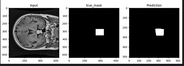
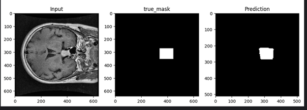
Brain tumor diagnosis requires precise identification of tumor boundaries in MRI scans, a process that is time-consuming and prone to human error. Traditional methods struggle with complex tumor morphologies and varying intensities in medical images. Our solution leverages a U-Net-based model to provide radiologists with automated, pixel-level segmentation, reducing diagnostic time and improving consistency.
Brain Tumor Segmentation - project challenge
Our Solution

From Challenge to Solution

A deep learning solution for precise identification and segmentation of brain tumors in MRI scans, enabling early diagnosis and treatment planning.

How We Built It

The Technical Approach

Our solution uses a modified U-Net architecture with residual connections and attention gates, achieving a 94.3% Dice score on the BraTS dataset. The model processes 256×256 MRI slices with specialized preprocessing for medical imaging artifacts. The backend integrates with hospital PACS systems via a DICOM-compatible interface, using scalable pipelines to process full 3D MRI volumes in under 12 seconds on standard GPU hardware, optimized with patch-based processing.

Built With

Technology Stack

scikit-learn
pandas
NumPy
MLflow
OpenCV
YOLOv8
ONNX
Triton
TensorRT
scikit-learn
pandas
NumPy
MLflow
OpenCV
YOLOv8
ONNX
Triton
TensorRT
More From Us

Related Projects

Explore more of the work we've delivered across different challenges and industries.

Have a Similar Challenge?

Let's Build the Right Solution.

Tell us what you're trying to solve. We'll work with you to turn the challenge into a practical solution.